The different examinations
Thyroid gland tumor follow-up examinations
are a focus of our competence center. An exact knowledge of the medical history and all prior findings are important to provide a high degree of safety. For your well-being and to ensure high-quality examinations, you will always be cared for by the same physician.
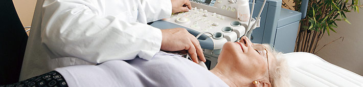
What is an ultrasound examination/sonography?
This painless examination makes it possible to evaluate the size and structure of the thyroid gland and other soft tissues of the neck. Nodules or inflammatory alterations can be detected.
A scintigraphic functional examination
• shows the metabolism of the thyroid gland while it is functioning. This makes it possible to distinguish hot (active)
• nodules from cold (inactive) nodules.
• shows whether active thyroid gland remnants are present after surgery.
• shows which nodules need not be subjected to a fine needle biopsy.
At the beginning of the examination you will be given a small quantity of a radionuclide (weak radioactive drug), which will be injected into a vein in your arm. After 12 minutes, a functional image of your thyroid gland will be recorded by a thyroid gland gamma camera.
An ultrasound-guided fine needle biopsy
is used in the cytological evaluation of nodules (benign/malignant) or unclear structural alterations of the thyroid gland (type of inflammation) by cytopathologist specialized in thyroid gland tissue examination.
A fine needle biopsy takes only a few seconds and is comparable to a blood-test. It is important to know that thyroid gland nodules are benign in more than 95% of all cases.
Blood tests
are used to determine thyroid hormones (hyperactive - hypoactive, fine adjustments), autoantibodies (autoimmune thyroid diseases), transport proteins and general parameters of inflammation (evaluation of viral inflammation over time, tumor markers after surgery) and blood coagulation values (before fine needle biopsy).